What is a platelet?
A platelet, or thrombocyte, is a non-nucleated structure which is derived from a mother cell, termed a megakaryocyte. The megakaryocyte is the largest structure in the bone marrow, with an abundant cytoplasm which is shed as small structures called platelets. Each megakaryocyte produces about 2000 platelets.
What is the function of platelets?
Platelets have a major function in coagulation, or clot production. Even though platelets are non-nucleated, they contain a sophisticated network of glycoproteins, receptors, and biochemistry, which help them to accomplish coagulation. When there is tissue injury, platelets are the first responders, adhering to tissue surface, changing shape and then aggregating to form a platelet plug. This series of reactions by platelets also calls into play the clotting factors, which subsequently go onto form the stable fibrin clot.
How do we recognzie normal platelets on the peripheral smear?
Normal platelets are 2.0-4.0 µm in diameter and appear discoid, pale blue and granular. The number of platelets in each microscopic field in a peripheral smear varies depending on the platelet count. However, there are normally about 13 – 20 platelets in a typical field when viewed using a 100x microscope objective.
How do we estimate the number of platelets on the peripheral smear?
Platelets are estimated under 100x magnification using a well-made and well-stained peripheral smear. Once the student finds a good counting area (100 red cells not touching) then they will move to a magnification of 100x. The student goes through 10 counting fields, clicking off the number of platelets seen in each area. A counter will be needed for this function. Then the student will average the count in the 10 fields he has counted and multiply by 20,000. This will provide a platelet estimate which can be compared to the reference range of platelets which equals 150-450 × 109/L platelets.
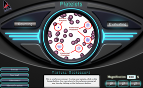
What are the three platelet abnormalities that can be visualized on the peripheral smear?
The three most common morphologic platelet abnormalities are giant platelets, platelets with abnormal granulation, and platelet fragments. Giant platelets are seen as structures which are increased in size well beyond 4 microns. At times giant platelets are as large as the red cell in the peripheral smear. In several of the bone marrow disorders, platelets are seen with no granulation, in which case they look like empty blue shells, or abnormal granulation, appearing scattered and larger throughout the platelet. The last abnormal feature is the megakaryocytic fragment, where large pieces of the megakaryocyte cytoplasm have disengaged from the cell and made their way into circulation.
What is a critical platelet level?
The reference range for the platelet is 150 × 109/L and 450 × 109/L. Platelet counts which are less than 30 × 109/L are in the critical range and the patient may be at risk for spontaneous bleeding. Evidence of decreased platelet counts may be noticeable nose bleeding, gum bleeding, small skin spots called petechiae, and excessive bruising or blotchiness called purpura.
 Have you ever cut your finger when slicing fruit or torn your knee after a gnarly fall? You can thank platelets for stopping you from bleeding out. Platelets are the cells that help form blood clots. Platelets start the clotting process by traveling to your wound and sticking to form a clump of cells. Sticking and clotting factors are based on the health and number of platelets you have. Your challenge is to look at platelets under a microscope, estimate how many platelets there are, and notice abnormal platelets.