What is the Gram staining (GS) procedure?
Developed by Hans Christian Gram in 1884, the Gram staining procedure is a simple stain of reagents and clinical specimens designed to help identify bacteria. As a differential stain, it groups most bacteria into 2 groups: gram-positive (GP) (distinguished by a blue color) and gram-negative (GN) (distinguished by a pink color). This division of bacteria is based on the chemical make-up and structure of the bacterial cell wall and its interaction with the reagents of the Gram staining procedure. Gram-positive bacteria have a thick peptidoglycan layer with excessive teichoic acid and cross linkages and contain a small amount of lipids, all of which aid in resisting decolorization. Gram-negative bacteria, on the other hand, have little peptidoglycan and more lipids, which lead to the cell being stained pink.
What are the reagents and their functions in the Gram Staining procedure?
The Gram stain reagents, listed as they are used in the staining procedure, are crystal violet, iodine, ethanol/acetone and safranin. The initial reaction of the staining procedure is with the addition of the primary stain, crystal violet, to the bacterial smear. Crystal violet colors the bacterial cell purple. Addition of the iodine reagent which is a mordant fixes the crystal violet to the cell wall. The crystal violet-iodine complex is attached more tightly to GP cell wall than the GN cell wall. The Ethanol/Acetone solution decolorizes or removes the crystal violet-iodine complex primarily from the GN cell wall. The GP cell wall will retain the crystal violet due to the presence of excessive teichoic acid and will remain purple. The counterstain or safranin is the final stain in the procedure. Safranin stains the colorless GN bacteria pink.
What are the sequential steps in a smear preparation?
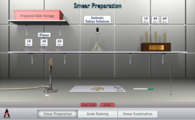
The source of the culture (broth or plate) will determine the steps in making the smear. For a plate culture, a very small drop of isotonic saline is placed in the center of the labeled slide. A small amount of culture is removed from the plate with the sterilized loop and placed on the drop of saline. This mixture is mixed and the final smear is about the size of a dime. For the broth, no saline is needed. The smear is prepared as for the plate culture. For both a plate culture and a broth culture, the smear is air-dried and heat-fixed before the GS procedure.
How do we evaluate the stained smear?
The stained smear is examined under 100x magnification. Several fields will be examined to determine the shape and Gram reaction-color of the bacteria. If the stain has been done correctly, the bacteria should be pink or purple. The GP bacteria will be purple, whereas the GN will be pink or reddish.
What are two critical steps in the GS procedure?
Decolorization is the most important step in the GS procedure. Decolorization may be impacted when excess washing or rinsing of the slide after the addition of crystal violet results in GP bacteria incorrectly staining red or pink. The length of time a stain is applied before it is rinsed also affects decolorization. Although 10 seconds is the typical time for decolorization, the time may vary depending on the source of the specimen (broth, saline, or sputum) as well as the thickness of the smear. Over-decolorization is evident when GP bacteria appear as pink upon microscopic examination; under-decolorization will result in GN bacteria to appearing as purple.
 A gram stained smear is a key diagnostic tool that aids in the preliminary diagnosis of infectious agents in a clinical microbiology laboratory. This differential stain is based on the cell wall differences of gram positive and gram negative bacteria. Gram stain can detect the presence of microorganisms in a patient specimen and also can be employed in identifying microbes from a culture. All clinically important bacteria can be identified using the gram stain with the exception of intracellular bacteria such as Chlamydia, those that lack cell wall such as Mycoplasma and those organisms that cannot be resolved by light microscopy such as spirochetes. The challenge is fourfold: To prepare a bacterial smear for Gram staining, to perform the Gram staining procedure, to identify the shape and gram reaction of the bacteria, and to evaluate the quality of the stain.


